Here are some of the interesting patients for the week. The photos of the patient are located below the history.
Patient 1: Female in her mid forties, referred to us for a new conjunctival lesion on the left eye in the setting of HIV. Presumed at outside to be a squamous cell carcinoma. On exam, vision in the left eye was 6/9 (about 20/30). Unfortunately as you can see below, the disease is already very advanced and has intraocular extension.
 |
| Squamous cell carcinoma. |

 |
| Note the peaking of the pupil toward the lesion (which is located inferotemporally in this left eye). This is a bad sign and indicates with near certainty that there has been intraocular extension. |
Patient 2: An 11 year-old boy who was playing last week when he had trauma to the right eye. Exam initially seemed unremarkable, except for a greyish spot on the superonasal aspect of the conjunctiva/sclera of the right eye, with mild surrounding injection. Upon dilated exam, there were no signs of infection, but there was a large, pointy mass protruding into the eye in the exact same location as the external spot. I looked, and looked again. I couldn't identify any retinal tears and couldn't exactly tell what the "pointy mass" was. I asked Dr. Pons to take a look. He said, "it almost looks like a thorn!" We then asked more history and were told that the boy was hit in the eye by a thorn and tried to pull it out. Well, folks it was the thorn that was protruding into the eye! The darkish area on the outside was the base of the thorn, covered by conjunctiva. Incredible. (Of note, there also seemed to be a small macular hole, responsible for the decrease in visual acuity. There is a retina specialist visiting in two weeks, and the boy is scheduled for vitrectomy/removal of foreign body.
 |
| Greyish area is the base of the thorn, covered by conjunctiva. |
Patient 3: A 23 year old female with severe glaucoma of the left eye. Her right optic nerve was healthy (0.2 cup:disc ratio) and her left eye was totally cupped out (0.99 cup). Diagnosis? (This is a classic presentation and should be a "diagnosis from the doorway").
 |
| Diagnosis? |
 |
| Answer: Sturge-Weber, one of the phakomatoses. Not hereditary. Note the dilated, tortuous episcleral veins. (See the picture above too. You can tell the left eye is much more hyperemic than the right). She also had the typical "tomato-ketchup" fundus OS. They eye also appeared slightly buphthalmic (see the first photo--it is subtle). |
Patient 4: My favorite history of the day. A 34 year-old female sent to us from Ubombo Sugar Hospital (many sugarcane farms have a clinic/hospital). The history on the referral letter reads, "Was injured in August with hot soft porridge that splashed into her left eye. Now unable to open eye, painful and tearing. Blurred vision." I have never had a patient get soft porridge in his/her eye. The poor lady was in so much pain (photo below). She was photophobic as well.
 |
| Central stromal edema with Descemet's folds. The pain was from superficial bullae. We diagnosed as a disciform keratitis, likely secondary to HSV and unrelated to the "soft porridge" incident. We are treating with dexachlor drops (dexamethasone-chloramphenicol) and oral Acyclovir. |
Patient 5: 23 year old female with vision of hand motions OD and OS. She was unable to give a good history, but it was apparent on exam that she had severe keratoconus. It was the most advanced keratoconus I have ever seen. Contact lenses will not work--she really needs a corneal transplant. (And also, contact lenses are really not available in Swaziland). Her cornea was so thin on the left, I fear she will perforate sooner rather than later. She was also a vigorous eye-rubber. She had evidence of stromal scarring secondary old hydrops.
 |
| Patient with SEVERE keratoconus OS > OD. Her best corrected vision is hand motions in both eyes. |
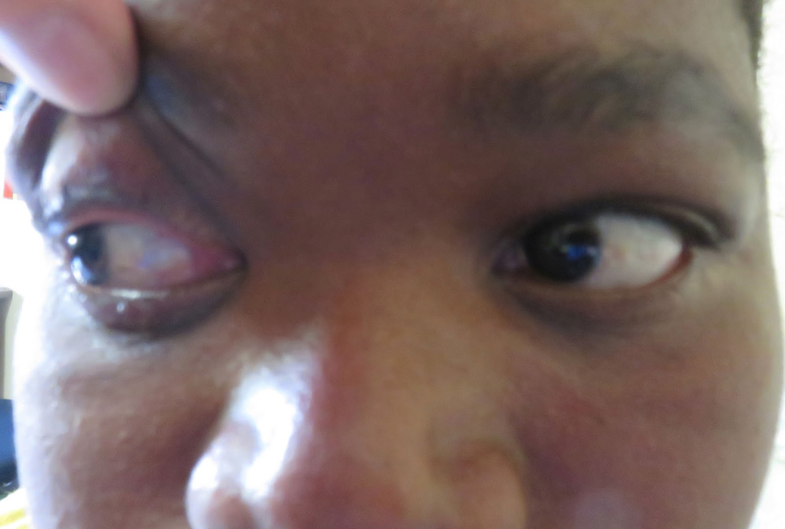
Patient 6: 25 year old female with acute onset of pupil-sparing third nerve palsy. She also had a headache. We took her blood pressure (elevated at about 190/100) and blood glucose (within normal limits). The etiology is likely microvascular (ischemic), but we referred her to a neurologist and she will likely get a scan (although head scans are difficult to obtain here). The nearest MRI is in South Africa.
 |
| Complete ptosis on the right. |
 |
| Primary gaze. Note the "down and out" position of the affected right eye. |
 |
| Right gaze |
 |
| Left gaze. |
 |
| Upgaze. |
 |
| Downgaze. |
Below: Welcome (an eye technician) does patient education prior to the start of Wednesday clinic. After he goes through all the posters at the front (on topics such as ocular trauma, HIV, etc), the patients are free to raise their hands and ask questions. My favorite question of the day (translated to English for me by Sister Senani): "If you have eye trauma, I have heard you are supposed to put salt in the eye. Is this true? " Some of the patients started laughing, but Sister insists that the patients will put all sorts of things in their eyes after trauma, from different herbal remedies, to salt, to urine!
 |
| Welcome doing ocular education with the patients prior to the start of Wednesday clinic at Good Shepherd Eye Clinic. |
 |
| Beautiful Swaziland countryside. |
No comments:
Post a Comment